

New paper in Nature Communications
A new laser-driven X-ray and proton micro-source
03.12.2020
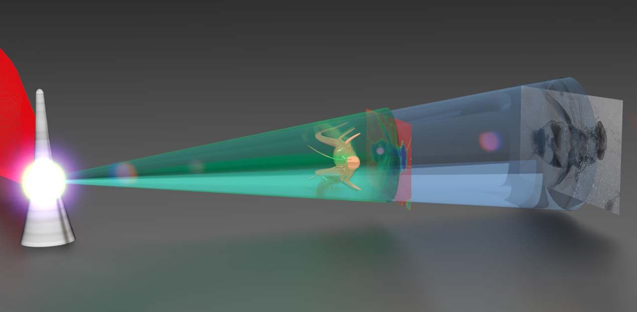
An international research team led by scientists around Prof. Katia Parodi and Prof. Jörg Schreiber from the Ludwig-Maximilians-University Munich has demonstrated a novel method to build a combined proton and X-ray microsource that enables high-resolution imaging, e.g., of biological samples, with both modalities at once.
Conventional radiography machines produce only a single species of radiation (i.e., protons or x-rays) and require significant post-processing steps to register images from different acquisitions on top of each other. This can proof difficult, especially if living (e.g., breathing) specimen or fast evolving processes are imaged. However, it is often required in order to leverage the complementary information contained in both modalities; for example, in the treatment planning procedures for modern charged particle therapy of cancer.
Laser-driven plasmas have long been known to emit a wide range of energetic radiation modalities including x-rays and protons. Here, the team used one of the most powerful lasers currently available, the Texas Petawatt laser at the University of Texas at Austin, and focused it to enormous intensities on a tiny needle with a width of just 1/20th that of a human hair and a pulse duration of 150 femtoseconds (that is 0.00000000000015 seconds). In such extreme conditions, electrons and protons can be accelerated from the needle-target and high energy photons (X-rays) can be generated.
The new trick here was the needle-target, which replaced a more conventional planar foil target. This allowed the team to geometrically separate desired proton and X-ray radiation fields from the driving high-intensity laser and from unwanted co-existing radiation (especially high energy electrons) that can easily harm imaged samples and/or contaminate the recorded image contrast. In addition, the target was found to produce proton and x-ray sources with spectral and spatial properties very well suited for high-resolution imaging (including X-ray phase contrast imaging).
This is the first time that actual biological and technological samples have been imaged with a simultaneous laser-driven X-ray and proton micro-source. However, the researchers note that their experiment marks just the first step in demonstrating the potential of such compact laser-driven multi-species micro-sources. In order to become widely applicable, the source and detector technology will require further improvements especially in particle-energy levels and repetition rate, which the researchers are confident to address with the next generation of high-repetition rate lasers currently coming online.
Future research will also investigate related applications of this type of source, e.g. in small sample test scenarios for image guided cancer therapy, e.g., using X-rays for imaging and simultaneously generated protons for irradiation.
Originally published on Attoworld. Text: Thorsten Naeser
Original Publication
- T. Ostermayr, C. Kreuzer, F. Englbrecht, J. Gebhard, J. Hartmann, A. Huebl, D. Haffa, P. Hilz, K. Parodi, J. Wenz, M. E.. Donovan, G. Dyer, E. Gaul, J. Gordon, M. Martinez, E. McCary, M. Spinks, G. Tiwari, B. M. Hegelich, and J. Schreiber, "Laser-driven x-ray and proton micro-source and application to simultaneous single-shot bi-modal radiographic imaging", Nature Communications 11, 6174 (2020)